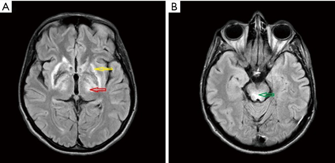
Figure 7.
Magnetic resonance imaging of the brain of a 23-year-old female with Wilson disease revealed a typical variety of MRI changes during the course of the disease. On FLAIR images solid hyperintensity changes in thalami (red arrow) and midbrain (green arrow), and modest hyperintense and hypointense signals in the putamen (yellow arrow) with markedly hyperintense signals in lateral putaminal margins are seen. Putaminal changes partly reflect the course of degeneration and are irreversible. MRI, magnetic resonance imaging; FLAIR, fluid attenuation inversion recovery.

