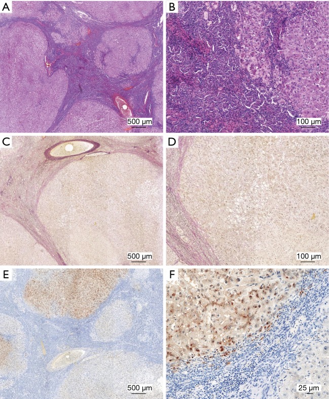
Figure 2.
Histochemical copper stains in diagnosis of Wilson’s disease. Liver specimens taken from a Wilson’s disease patient were stained with hematoxylin & eosin (H&E) (A,B), Verhoeff’s Van Gieson stain (EVG) (C,D), or subjected to rhodanine stain (E,F). In rhodanine stain, nuclei stain blue, while positive granules red or brown in color reflect cytoplasmic accumulation of copper. Please note the large number of inflammatory cells within the tissue. Bridging fibrosis and cirrhosis is seen in H & E stain. Collagen fibers in EVG stain appear red, while elastic fibers stain blue to black. Images were taken at different magnifications. Space bars represent 500, 100 or 25 µm, respectively. The permission to analyze anonymized human liver samples is covered by an ethical vote (EK 186/15) from the Institutional Ethics Review Board of the Medical Faculty at the RWTH University Hospital Aachen.

