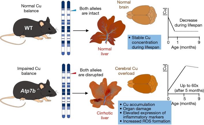
Figure 5.
Atp7b null mice, a well-established experimental model of Wilson’s disease. The mouse homolog of the Wilson’s disease gene (Atp7b) is located on mouse chromosome 8 in close proximity of the microsatellite marker D8Mit3 (45). Homozygous disruption of the Atp7b gene in mice results in a phenotype resembling human Wilson’s disease (43). In the life span of respective mice, two distinct phases with respect to the Cu balance are distinguishable. During weaning, progeny of homozygous mutant females shows a significant deficiency in Cu at birth in comparison to wild-type mice. This is due to the fact that milk from the mutant glands is Cu deficient. Thereafter, Atp7b null mice display a gradual accumulation of Cu in various organs that increase in liver up to a level 60-fold greater than normal. This intoxication induces the generation of cirrhotic livers and neuronal abnormalities. In contrast, wild-type mice show a decrease in copper content during the first month of life to a level that remains stable for the rest of lifespan.

