. 2019 Apr 23;10(19):5114–5123. doi: 10.1039/c9sc00591a

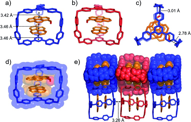
Fig. 4. (a) Side-on stick diagrams view of the crystal structures of (PCA)2AzaEx2Cage6+ forms with different chirality including (a) R (blue) and (b) S (red). The axial chirality of the complex results from the different relative rotation directions of the carbonyl groups in the two PCA guests within the cage cavity. The interplane distances between the platforms in the host and the two guests are 3.42, 3.46, and 3.46 Å, indicating the occurrence of π–π stacking interactions. (c) Top view of the X-ray crystal structure of (PCA)2AzaEx2Cage6+. The distances between the two carbonyl oxygen atoms and the corresponding BIPY2+ pillars are 2.78 and 3.01 Å, respectively, indicating the occurrence of dipole–cation or dipole–dipole interactions. (d) Side-on stick diagram view overlaid with a space-filling representation of the crystal structure of (PCA)2AzaEx2Cage6+. (e) Side view of the crystal lattice of (PCA)2AzaEx2Cage·6PF6 showing that complexes of different chirality undergo stacking with each other, making the crystal lattice a racemic mixture. PF6 counterions are omitted for the sake of clarity.

Fig. 4