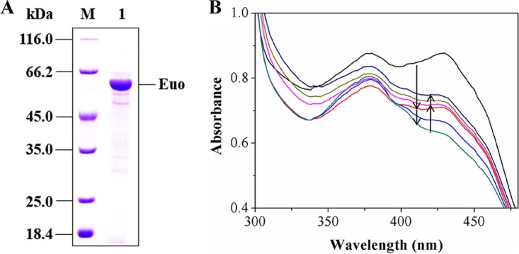
FIG 4.

(A) SDS-PAGE analysis of the purified recombinant Euo. M, protein marker; 1, purified protein. (B) Changes in UV-visible absorption spectra during the reduction of CoQ1 with reduced EtfAB-I by Euo coupling with the reduction of EtfAB-I with pseudooxynicotine by Pno. Initially, the reaction mixture contained 50 mM Tris-HCl (pH 8.5), 1 mM pseudooxynicotine, 0.05 μM purified Pno, and 30 μM purified Euo (black line). The mixture was then treated by adding 8 μM purified EtfAB-I (red line). The reduction of Euo proceeded with time (light-blue and dark-green lines). Upon stabilization of the spectrum, 32 μM CoQ1 was added and Euo was reoxidized (pink line). The reoxidization proceeded with time (light-green and dark-blue lines). The downward arrows indicate the reduction of Euo after the addition of EtfAB-I, whereas the upward arrows indicate the reoxidation of Euo after the addition of CoQ1. Neither oxidized nor reduced CoQ1 has absorption in the wavelength window. The time between the first two records (between the black line and dark-blue line) was 45 s. The rest of the records were done every 45 s.
