FIG 4.
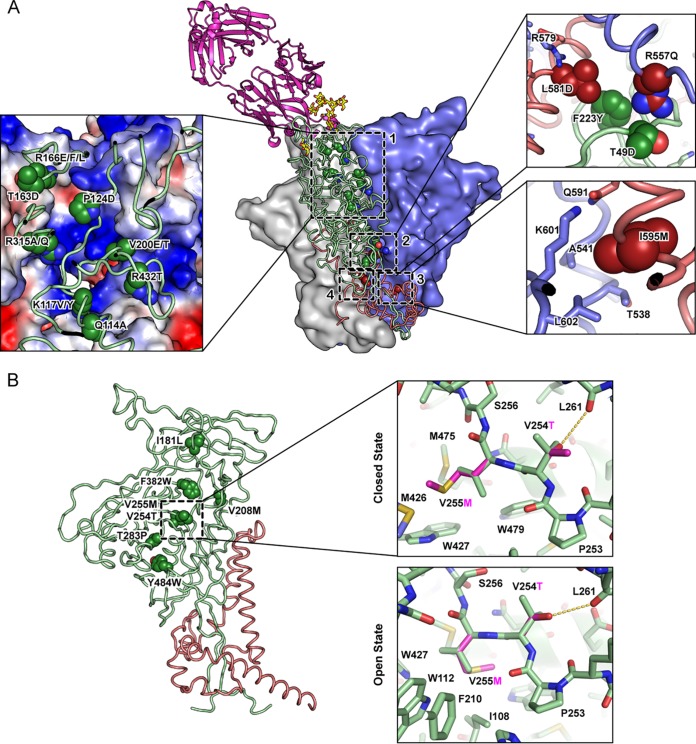
Neutralization of the electropositive apical cavity and increased packing at the inner-outer domain interface increase presentation of Env in a PG16-recognized conformation. From the mutational scan of gp160BaL and gp140DU422, substitutions were identified and validated that enhance PG16 binding. (A) Mutations at subunit interfaces clustered to four sites, shown on a structural model of closed EnvBaL. PG16 is magenta, interacting glycans are yellow, two Env protomers are shown as gray and blue surfaces, and the third Env protomer is shown as green (gp120) and brick red (gp41) ribbons. In the magnified inset of site 1, the electrostatic potential on the surface of two Env protomers is plotted from positive (blue) to negative (red). Previously described mutation L544Y is found at site 4. (B) Mutations in the core generally increased hydrophobic packing along the inner-outer domain interface of gp120. In the magnified insets, V255M (magenta) fills a void in the closed conformation, whereas modeling predicts V255M has steric clashes in the open conformation, with only a single methionine rotamer being accommodated.

