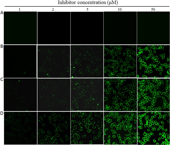
FIG 4.
Visualization of lipopeptide inhibitors bound to the target cell membrane. Different concentrations of FITC-labeled P-52 (A), LP-52 (B), LP-80 (C), and LP-83 (D) were preincubated with TZM-b1 cells for 30 min, followed by washes, and the fluorescence intensities of membrane-attached inhibitors were observed under a confocal microscope.

