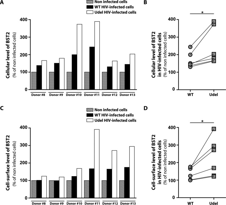
FIG 4.
Cellular and cell surface expressions of BST2 in infected macrophages. MDMs from different donors were infected with VSV-G-pseudotyped WT NL4-3 (WT) or Vpu-deficient NL4-3 (Udel) HIV-1. Five days later, the intracellular (A and B) and cell surface (C and D) levels of BST2 in noninfected or VSV-G-pseudotyped WT NL4-3 (WT) or Vpu-deficient NL4-3 (Udel) HIV-1-infected MDMs were analyzed by flow cytometry using anti-BST2 and anti-CAp24 antibodies. Mean fluorescence intensity (MFI) values for BST2 in CAp24-positive cells were normalized to that for noninfected cells. For panels B and D, each symbol represents the normalized BST2 MFI values of MDMs for each donor. Statistical significance was determined using unpaired two-tailed Student’s t test (means ± SEM; n = 3 donors). *, P ≤ 0.05.

