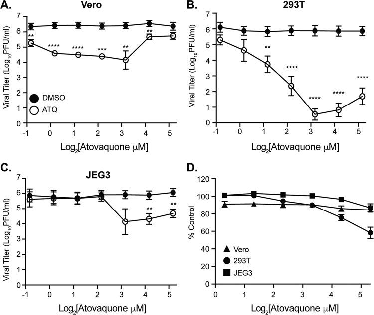
FIG 2.
Atovaquone inhibits ZIKV infectious virion production in mammalian cells. (A to C) Vero (A), 293T (B), and JEG3 (C) cells were infected with ZIKV MR766 at an MOI of 0.1 in the presence of atovaquone (open symbols) or DMSO control (closed symbols). Virus-containing supernatants were collected at 36 h postinfection, and infectious virus was quantified by plaque assay on Vero cells. The mean and standard error of the mean (SEM) are shown. For Vero and JEG3 cells, data represent three independent experiments, each with internal triplicates; for 293T cells, data represent four independent experiments, each with internal technical triplicates (**, P < 0.01; ***, P < 0.005; ****, P < 0.0001 [Student’s t test]). (D) Cells were incubated with increasing concentrations of atovaquone or DMSO control for 36 h and subsequently stained with Sytox green reagent to visual dye-permeable dead cells. Dead (Sytox-positive) cells were quantified by microscopy on a CellInsight CX7 high-throughput platform. Data are represented as percentage of alive cells (Sytox negative) compared to those in the DMSO control. The mean and SEM are shown. Data represent three independent experiments, each with internal technical triplicates.

