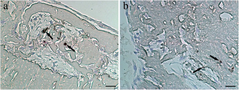
Fig. 5.

Immunohistochemical staining of CD34 in PDCs-CGF complex (magnification, × 400). PDCs were dispersedly distributed among the CGF fibrin matrix. CD34-positive cells (arrow) are visible (a, b). Scale bar = 100 μm. CD cluster of differentiation, PDCs periosteum-derived cells, CGF concentrated growth factor. All experiments were performed independently (each experiment used PDCs and CGF from a different rabbit) and repeated three times
