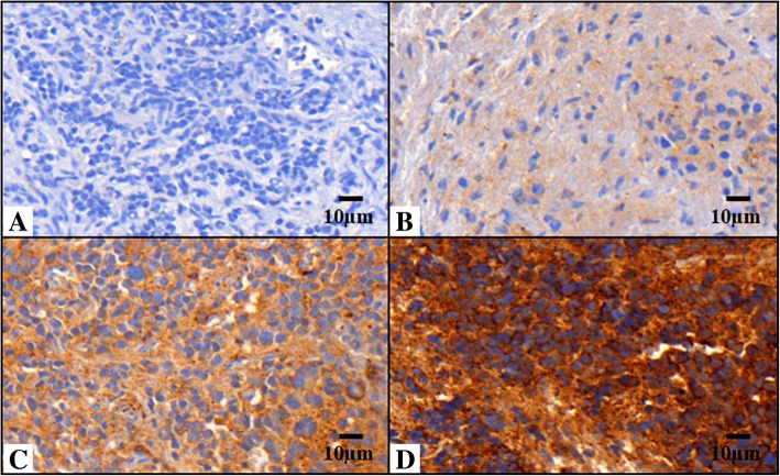
Fig. 1.
Vitronectin pattern in neuroblastic tumors. Images immunostained with antibody anti-vitronectin (VN) at 40X. a Sample corresponding to negative VN. b and c Samples corresponding to weak to moderate VN expression and ECM distribution only (defined as interterritorial VN). d Sample with strong VN expression with pericellular and intracellular location (defined as territorial VN)

