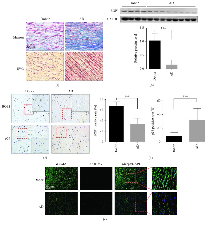
Figure 1.
BOP1 expression is decreased in ASMCs of AD patients. (a) Images of Masson staining showed collagen (blue) and muscle fibre (red) in the aortic media derived from AD patients and donors (upper panel). Representative images of EVG staining indicated the broken elastic fibre in aortic samples derived from AD patients and donors (lower panel). (b) BOP1 protein expression in the aortic media of donors (n = 4) and AD patients (n = 8) was detected by western blotting, and the related expression level was detected by statistical analysis and shown. (c) Representative image of the aortic specimens stained by BOP1 and p53 by performing IHC. (d) The positive rate was detected by statistical analysis and shown. (e) The 8-hydroxy-2′-deoxyguanosine (8-OHdG) level in the aortic media tissues were detected by performing immunofluorescence and the representative images are shown. Scale bar 50 μm. Data are presented as mean ± SD. ∗∗∗ P < 0.001 determined by Student's t-test.

