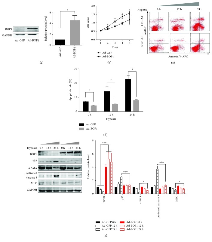
Figure 2.
Overexpression of BOP1 attenuated HASMC apoptosis under serum-free and hypoxia condition. (a) The expression of BOP1 in HASMCs was detected after it had been infected with Ad-BOP1 or Ad-GFP for 24 h (left panel). Statistical analysis is shown (right panel). (b) HASMCs were infected with Ad-BOP1 or Ad-GFP for 24 h. The cells were suspended and reseeded in 96-well plates. CCK-8 assays were performed to assess the influence of BOP1 on HASMC proliferative ability. The growth curve is shown. (c) After being infected with Ad-BOP1 or Ad-GFP for 24 h, HASMCs were administrated in serum-free and hypoxia condition for the time shown. Apoptosis was detected by Annexin V-APC/7-AAD staining and flow cytometry followed. The representative images are shown. (d) The statistical analysis of apoptosis rate is shown. (e) Western blotting was performed to detect the BOP1, p53, α-SMA, activated caspase 3, and MLC expression. The representative image is shown (left panel). The statistical analysis is shown (right panel). Data are representative of three independent experiments and presented as mean ± SD. ∗ P < 0.05, ∗∗ P < 0.01, and ∗∗∗ P < 0.001 determined by one-way ANOVA.

