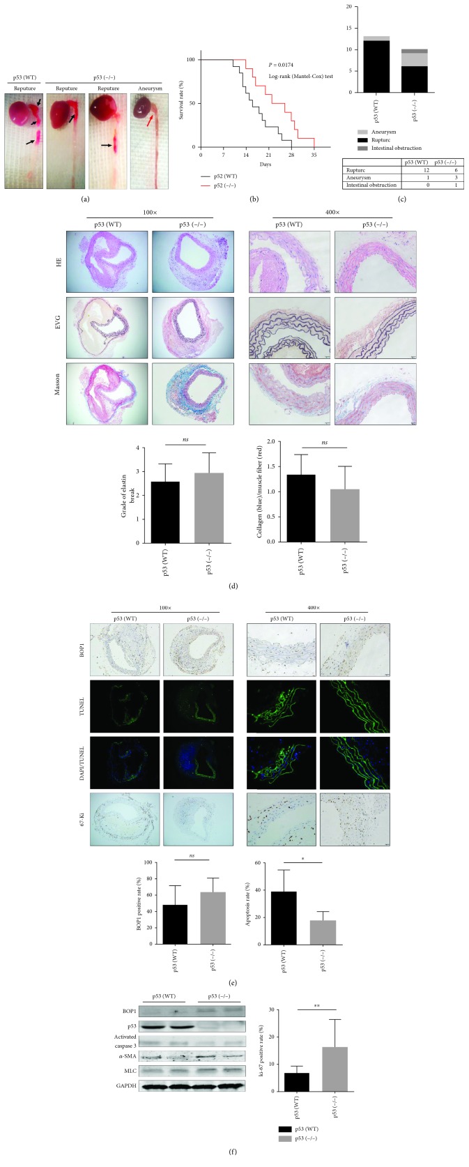
Figure 6.
Knockout of p53 reduced the occurrence of AD in mice. (a) Representative images of gross aortic samples are shown. (b) Kaplan-Meier survival curve is shown. (c) The death reason is summarized and shown. (d) Representative staining of aorta sections with HE, Masson, and EVG. Graphs show semiquantification of elastic fibre broken grade and collagen/muscle fibre ratio. (e) Representative images of the aortas performed with TUNEL assays, IHC staining with anti-BOP1 antibody and anti-ki-67 antibody. The positive rate is shown (right panels). (f) Western blotting was performed to detect the BOP1, p53, activated caspase 3, α-SMA, and MLC expression of the aortas. Data are presented as mean ± SD; ns: no statistical significance; ∗ P < 0.05, ∗∗ P < 0.01, and ∗∗∗ P < 0.001 determined by one-way ANOVA.

