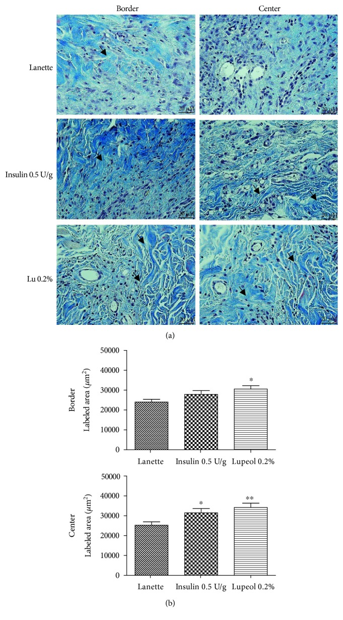
Figure 4.
Masson's trichrome-stained skin tissue sections on day 14 postwound induction in streptozotocin-induced hyperglycemic rats (a). Labeled area of total collagen fibers (b) (μm2) in the border and central region of rats' hyperglycemic wounds treated with Lanette, insulin 0.5 U/g, or lupeol 0.2% for 14 days. ∗ p < 0.05 and ∗∗ p < 0.01 vs. Lanette group, using ANOVA followed by the Newman-Keuls test. Bar represents 20 μm. Black arrows indicate the presence of total collagen fibers. Lu 0.2% = lupeol 0.2%.

