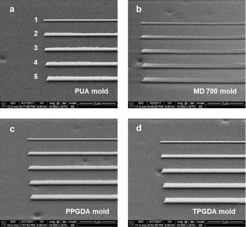
Fig. 3.

SEM images of replicated UV resin molds by UV-NIL from a Si master. Nanochannel depth increases from top to bottom as indicated by number in the image. The numbers 1–5 correspond to nanochannels 1–5 in Fig. 2. For low surface energy resins with high monomer molecular weight (PUA and MD 700), saw-tooth defects are seen for the replicated nanoprotrusions from deeper nanochannels due to uneven and incomplete resin filling. The SEM images were taken with a tilt angle of 52°.
