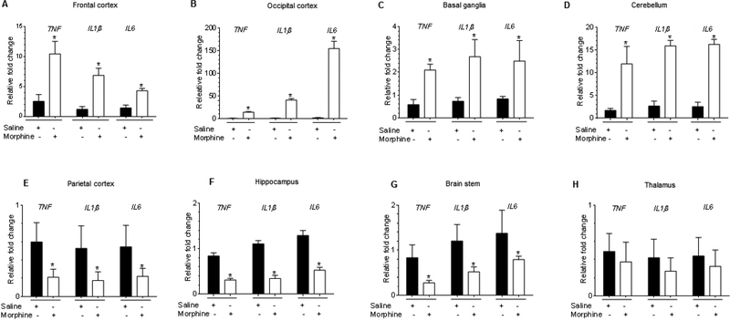
Figure 7.
Morphine-dependent rhesus macaques exert brain region-specific upregulation of proinflammatory cytokines. (A-H) qPCR demonstrating the relative expression of proinflammatory cytokines such as TNF, IL1β, and IL6 in frontal cortex (A), occipital cortex (B), basal ganglia (C), cerebellum (D), parietal cortex (E), hippocampus (F), brain stem (G), and thalamus (H) of morphine-dependent rhesus macaques. GAPDH was used as a loading control for mRNA expression of cytokines. Data are presented as mean ± SEM; n = 4. Abbreviations: S: Saline administered macaques, M: Morphine administered macaques, GCL: Granular cell layer, WM: White matter. Student t test was used to determine the statistical significance: *, P<0.05 vs. saline.

