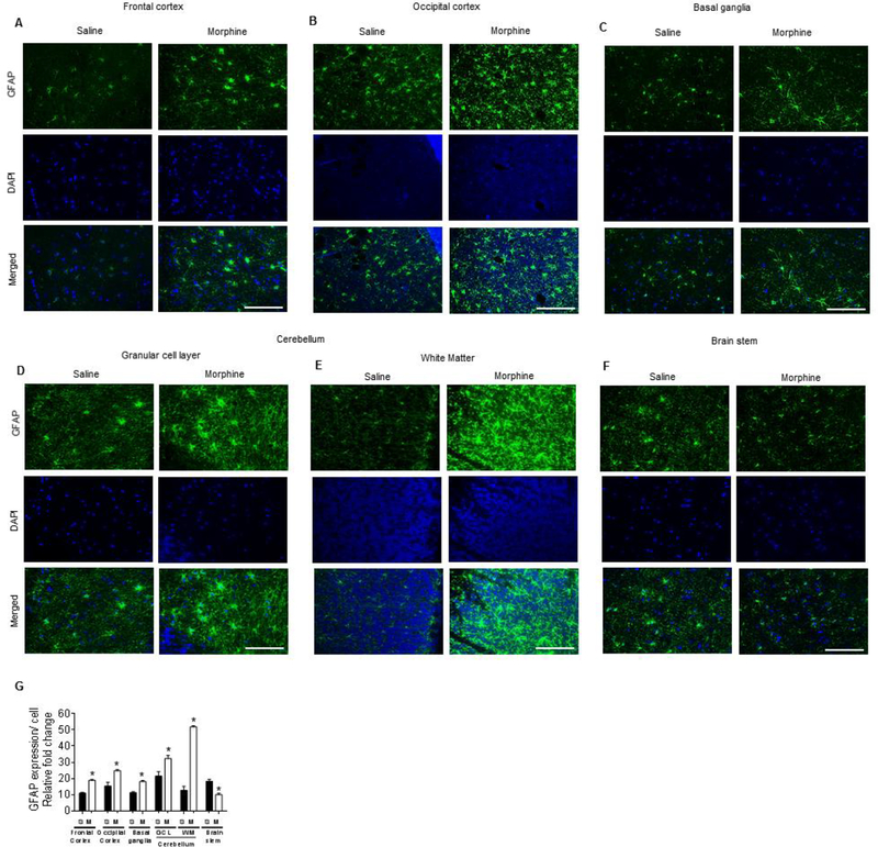
Figure 8.
Morphine-dependent rhesus macaques exert brain region-specific activation of astrocytes. A-F) Representative immunohistochemistry images showing the expression of GFAP in frontal cortex (A), occipital cortex (B), basal ganglia (C), different layers of cerebellum – granular layer (D) and white matter (E), and brain stem (F) of morphine-dependent rhesus macaques. Scale bar: 50 μM. (G) Densitometric analysis of GFAP positive cells using ImageJ software in the frontal cortex, occipital cortex, basal ganglia, different layers of cerebellum— granular layer and white matter, and brainstem of morphine-dependent rhesus macaques. Data are presented as mean ± SEM; n = 4. Student t test was used to determine the statistical significance: *, P<0.05 vs. saline.

