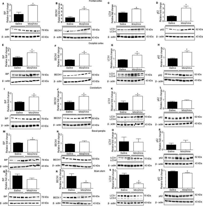
Figure 9.
Morphine-dependent rhesus macaques exert brain region-specific activation of astrocytes through ER stress with defective autophagy. (A-D) Representative western blots showing the protein levels of BiP (A), BECN1 (B), LC3-II (C) and p62 (D) in the frontal cortex of morphine-dependent and saline administered rhesus macaques. (E-H) Representative western blots showing the protein levels of BiP (E), BECN1 (F), LC3-II (G) and p62 (H) in the occipital cortex of morphine-dependent and saline administered rhesus macaques. (I-L) Representative western blots showing protein levels of BiP (I), BECN1 (J), LC3-II (K) and p62 (L) in the cerebellum of morphine-dependent and saline administered rhesus macaques. (M-P) Representative western blots showing the protein levels of BiP (M), BECN1 (N), LC3-II (O) and p62 (P) in the basal ganglia of morphine-dependent and saline administered rhesus macaques. (Q-T) Representative western blots showing the protein levels of BiP (Q), BECN1 (R), LC3-II (S) and p62 (T) in the brain stem of morphine-dependent and saline administered rhesus macaques. β-actin was used as a loading control for all experiments. Data are presented as mean ± SEM; each group (n = 4). Student’s t-test was used to determine the statistical significance: *, P<0.05 vs. saline.

