
Figure 1. Cardiac RhoE deficiency induces excessive post-MI inflammation.
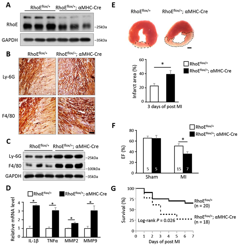
(A) Immunoblot for RhoE in 3 pairs of RhoEflox/+ and RhoEflox/+;αMHC-Cre mouse hearts. (B) IHC staining for neutrophil marker (Ly-6G) and macrophage marker (F4/80) in RhoEflox/+ and RhoEflox/+;αMHC-Cre mouse hearts on day 3 post MI. Scale bar: 0.2 mm. (C) Immunoblot for Ly-6G and F4/80 in whole heart lysates from the above mice. (D) qRT-PCR for IL-1β, TNFα, MMP2 and MMP9 in whole heart lysates from above mice. (E) TTC staining for infarct area (top panel, outlined by dashed line). Scale bar: 1.0 mm. Infarct area was quantified to left ventricle area in 3 pairs of mice (bottom panel). (F) Ejection fraction of sham and MI mice on day 3 post MI. (G) Kaplan-Meier survival curves of mice in the first week post MI. n: number of mice. *: P < 0.05.