Figure 4.

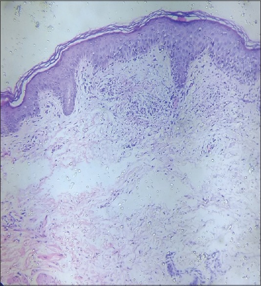
Histopathology showing hyperkeratosis, irregular acanthosis, focal hydropic degeneration in the basal layer. Dermal perivascular mononuclear cell infiltrate with fibrinoid deposits

Histopathology showing hyperkeratosis, irregular acanthosis, focal hydropic degeneration in the basal layer. Dermal perivascular mononuclear cell infiltrate with fibrinoid deposits