FIGURE 6.
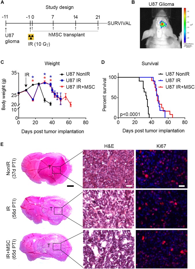
hMSC administration does not compromise survival of glioma-bearing mice. (A) Schematic representation of the study design. U87 glioma cells were intracranially transplanted into the striatum of nude mice. After 10 days, mice received whole-brain radiation (total dose of 10 Gy) and the day after, 5 × 10⁵ hMSCs were administered intranasally once per week for 4 weeks and time of death was monitored. (B) Xenolight DiR-labeled U87 glioma cells were locally transplanted into the striatum of nude mice. Images show bioluminescence activity in a representative animal 24 h after cell transplantation. (C) Animal weight was measured during the course of the experiment. Note the weight loss after radiation exposure. (D) Kaplan–Meier curve showing the percentage of survival of glioma-bearing mice. Note that IR and IR+MSC mice exhibited similar response, increasing survival as compared to NonIR mice. (E) Histological images of brain tumors at the time of death (indicated as days post tumor implantation, PTI) in the last individual surviving for each experimental group. H&E: Hematoxylin and eosin staining. Scale bar 1 mm (25 μm for details). Data are represented as mean ± SEM. n = 11–12 per group. ∗p < 0.0001 compared to U87 NonIR mice; Multiple t-test (C), Log-rank test (D).

