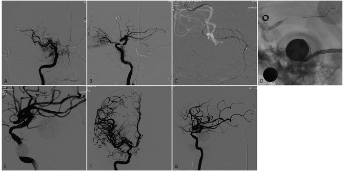
Figure 2.
A patient who had a previous history stroke and presented again with recurrent TIAs recently. (A,B) Right internal carotid arteriogram on anteroposterior and lateral view demonstrated a focal severe (90%) stenosis of the right supraclinoid ICA with flow reduction in the distal territory. (C) A 0.014 inch microwire was anchored in the distal branch of the MCA with the microwire tip manipulated a U- shaped loop inside the vessel. (D) The stenotic lesion was dilated with a 3.5 × 9-mm Gateway balloon. (E) The lesion showed significant improvement of the stenosis immediately after angioplasty. (F) and G. A 4.5 × 15-mm Neuroform EZ stent was deployed across the lesion, Final angiography showed near 20% residual stenosis with flow augmentation in the distal territory.

