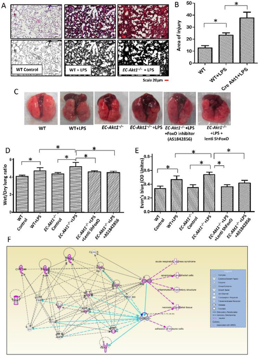
FIGURE 3: Endothelial Akt1 loss exacerbates LPS-induced ALI in EC-Akt1−/− mice, which is partially rescued by FoxO1/3a inhibition.
(A-B) Representative H&E images and bar graph showing the area of injury in the mouse lung sections indicating enhanced LPS induced ALI in EC-Akt1−/− mice compared to WT mice (n=6). (C) Representative lung images showing enhanced lung vascular leak (Evans blue extravasation) in EC-Akt1−/− mice, which were rescued by pulmonary FoxO1/3a inhibition using LentiShFoxO1/3a or FoxO1 inhibitor (AS1842856) i.p. 30mg/kg. (D) Bar graph showing lung wet/dry weight ratio as a measure of lung edema in LPS injured EC-Akt1−/− mice compared to WT mice and its partial reversal with pulmonary FoxO1/3a inhibition using LentiShFoxO1/3a or FoxO1 inhibitor (AS1842856) i.p. 30mg/kg (n=6). (E) Bar graph showing Evans blue extravasation confirming enhanced lung vascular leakage in EC-Akt1−/− mice and its partial rescue with FoxO inhibition using LentiShFoxO1/3a or FoxO1 inhibitor (AS1842856) i.p. 30mg/kg (n=6). (F) FoxO1/3a regulates ARDS genes and pathology in humans and rodents as identified from genome-wide association studies using ingenuity pathway analysis (IPA). * (p<0.05); # (p<0.01), One-way ANOVA-Ordinary for more than two groups (GraphPad Prism 6.01). Green lines indicate direct interaction with concerned pathway; Pink lines indicate indirect interaction with the concerned pathway; Blue lines indicate the interaction between genes of concerned pathway.

