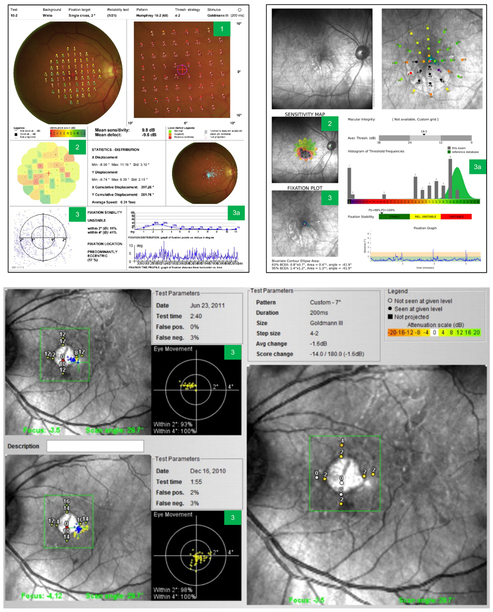
Fig. 2. Test outputs and how to read a microperimetry report.
Sample microperimetry reports from Nidek MP-1 (top left), MAIA (top right), and Optos OCT/SLO (bottom). Test outputs: 1) local defect map; 2) interpolation map; 3) fixation points; 3a) assessing fixation stability. The Optos microperimetry output shows progression data over time between 2 visits. MAIA, Macular Integrity Assessment; OCT, optical coherence tomography; SLO, scanning laser ophthalmoscope.

