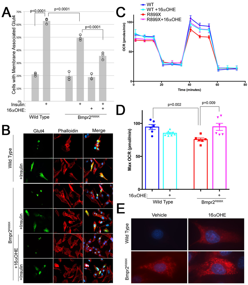
Figure 6 –
(A) Insulin mobilization of GLUT4 in mPMVEC is decreased in BMPR2R899X mice, and decreased further by pretreatment with 16αOHE1. Insulin mobilization was determined by counting perinuclear vs membrane associated GFP-tagged GLUT4 in each of 100 cells in each of three technical replicates (% in each replicate indicated by circles). Every group has three technical replicates: some are overlapping. Error bars are standard deviation; differences are significant at p<0.0001 by ANOVA, with comparisons indicated by Fisher’s LSD. Differences are also significant at p=0.01 by Wilcoxon non-parametric test. (B) Live cell images of mPMVEC transiently transfected with GFP-tagged Glut4, untreated and pretreated with 16αOHE1, either with vehicle or 15 minutes after treatment with insulin (which ought to mobilize Glut4 to the cell surface). (C) Seahorse Extracellular Flux analysis using the Mito Stress Protocol shows an increase in maximum oxygen consumption rate with FCCP treatment (@~40–60 minutes). (D) Quantitation of maximum oxygen consumption rate from data in (C). (E) A Mitosox Red assay suggests that the increased oxygen consumption in Bmpr2 mutant cells with 16αOHE1 is driven by increased mitochondrial superoxide production.

