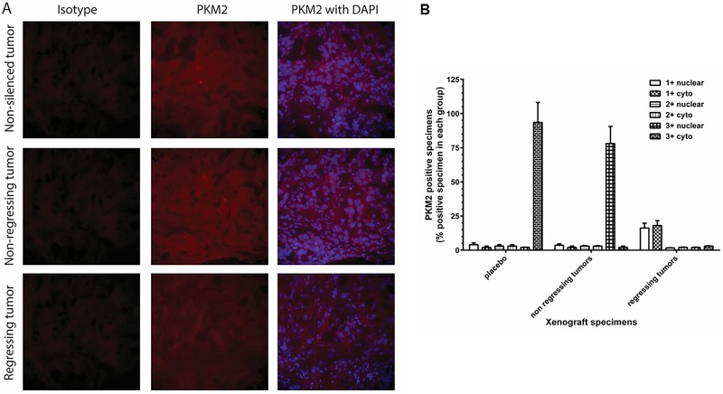
Fig 7. PKM2 expression in NSCLC tumor specimens.
(A) H358 cells (non-silenced, mock-transfected and shRNA-PKM2) were implanted subcutaneously in nude mice and the tumor growth of each mouse was monitored. The mice were euthanized on day 80 and the s.c. tumors were surgically excised. The tumor specimens were fixed with 4% paraformaldehyde, embedded in OCT, cryo-sectioned, and immunostained with anti-PKM2 antibody. PKM2 was stained with Alexa 594 (red) and DAPI was used for nuclear staining (blue). The sections were viewed in Nikon fluorescence microscope at 200 x magnification. (B) Extent of PKM2 immunostaining in cytoplasm and nucleus was evaluated as ≤1+ negative; 2+ positive; 3+ strongly positive. % positive of total number of tumor cells was shown as mean ± SD.

