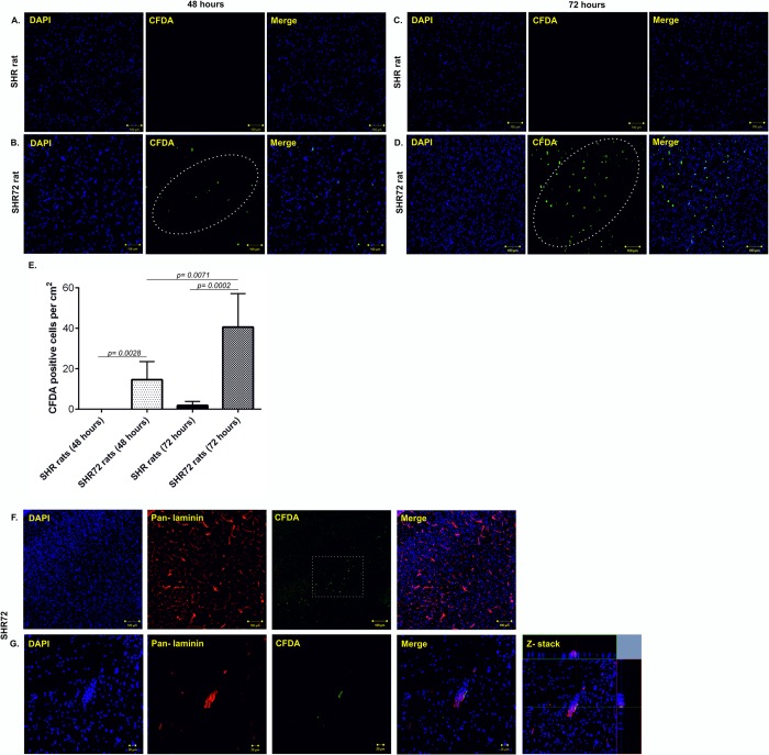
Fig 4. Active infiltration of CFDA—labeled PB-MoM cells into the area with neurofibrillary pathology.
(A) CFDA- labeled PB-MoM cells in control SHR brain tissue (brainstem) after 48 hours. (B) CFDA- labeled PB-MoM cells in transgenic SHR-72 brain tissue (brainstem) after 48 hours. (C) CFDA- labeled PB-MoM cells in control SHR brain tissue after 72 hours. (D) CFDA- labeled PB-MoM cells in transgenic SHR-72 brain tissue after 72 hours. (E) Quantification of CFDA- labeled PB-MoM cells in the brainstem of control and transgenic rats after 48 and 72 hours. (F, G) Double immunostaining with pan-laminin showed the presence of intravascular CFDA- labeled cells. Scale bar: 100 μm and 20 μm, n = 4.

