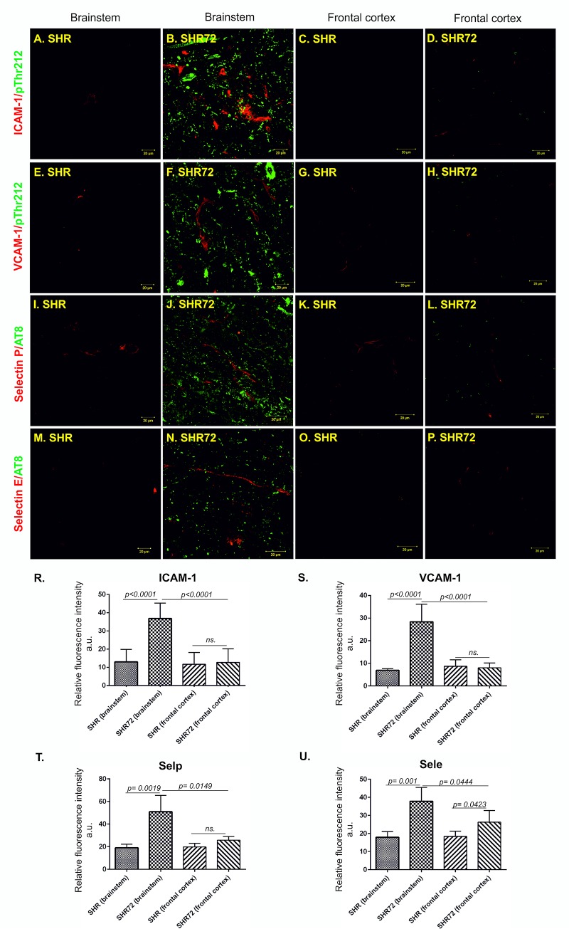
Fig 6. Expression of adhesion molecules is localized in the brain area affected by neurofibrillary pathology.
Immunostaining of adhesion proteins ICAM-1, VCAM-1 and selectins in transgenic and control rats. Confocal microscopy showed that brain capillaries stained with ICAM-1, VCAM-1 and selectins antibodies (red color) were distributed throughout the brain affected by neurofibrillary lesions immunolabelled with pThr212 and pSer202/pThr205 (AT8, green color). Low or no signal was detected in the brainstem of control rats and frontal cortex of control and transgenic rats. (A) Presence of ICAM-1 in the brainstem of a control subject, (B) increased expression of ICAM-1 in the brainstem of transgenic rats, (C) presence of ICAM-1 in the frontal cortex of control subject, (D) expression of ICAM-1 in the frontal cortex of transgenic rats. (R) Quantification of ICAM-1 expression. (E) Presence of VCAM-1 in the brainstem of control animal, (F) in the brainstem of the transgenic animal, (G) in the frontal cortex of control animals and (H) in the frontal cortex of transgenic rats (S) with the following quantification. (I) Presence of Selp in the brainstem of controls and (J) in the brainstem of transgenic rats. (K) Presence of selp in the frontal cortex of control and (L) transgenic rats. (T) Quantification of selp immunoreactivity in control and transgenic animals. (M) Presence of Sele in the brainstem of controls and (N) transgenic rats. (O) Presence of Sele in the frontal cortex of controls and (P) transgenic animals. (U) Quantification of Sele immunoreactivity in control and transgenic animals. Scale bar 20 μm, n = 10.

