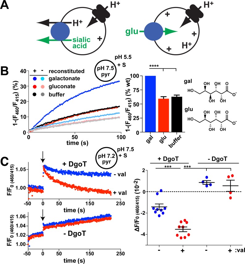
Fig 1. Coupled D-gal:H+ cotransport by E. coli DgoT.
(A) Diagram illustrating transport by sialin (left) and a VGLUT (right). A vacuolar-type H+ pump provides the H+ electrochemical driving force for both activities, but sialin uses the pH gradient to drive electroneutral cotransport of sialic acid with H+ out of the lysosome whereas the VGLUTs rely on membrane potential to drive glutamate uptake by synaptic vesicles against a pH gradient. (B, C) Liposomes containing pyr at pH 7.5 were reconstituted with (+) or without (−) E. coli DgoT and added to reaction buffer at pH 5.5. gal, glu (both 10 mM), or buffer alone were added at t = 0. The fluorescence emission at 510 nm with excitation at 460 nm was normalized to excitation at 415 nm (the isosbestic point for pyr versus pH). (B) The representative change in fluorescence ratio shows that D-gal but not glu or buffer control reduce the lumenal pH of proteoliposomes reconstituted with DgoT but not empty liposomes. Bar graph (right) shows the average rate of fluorescence change for glu and buffer relative to gal (n = 6). ****p < 0.0001. (C) Traces (left) show representative fluorescence ratios of proteoliposomes with (above) and without DgoT (below), both formed at pH 7.2, before and after the addition of 10 mM D-gal (at arrow) in external solution at pH 7.5, with or without 0.2 μM val. Both internal and external solutions contained 5 mM K+. Scatterplot (right) shows the difference of fluorescence decay between the addition of gal (t = 0) and the end of recording (t ≈ 220). ***p < 0.001 by one-way ANOVA with Bonferroni correction. n = 9 for proteoliposomes with DgoT; n = 4 for liposomes (see S1 Fig). The numerical data underlying this figure are included in S1 Data. DgoT, D-galactonate transporter; gal, galactonate; glu, gluconate; pyr, pyranine; val, valinomycin; VGLUT, vesicular glutamate transporter; WT, wild type.

