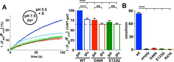
Fig 3. Functional residues coupled to proton or substrate transport.
(A) Representative fluorescence of proteoliposomes reconstituted with WT (blue) or mutant DgoT (D460N red, E133Q green) in response to addition of D-gal (darker color) or glu (lighter color) at t = 0 seconds. Bar graph (right) shows the average change in fluorescence at t = 90 seconds. Filled bars indicate galactonate, open bars gluconate (n = 11). ***p < 0.001; ****p < 0.0001. (B) Whole-cell uptake of radiolabeled 14C-galactonate by WT and mutant DgoT exogenously expressed in an E. coli DgoT knock-out strain. WT but not D46N or E133Q DgoT confer 14C galactonate uptake (n = 3). The numerical data underlying this figure are included in S1 Data. DgoT, D-galactonate transporter; gal, galactonate; glu, gluconate; ns, not significant; pyr, pyranine; WT, wild type.

