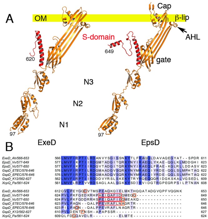
Fig 5. Domain arrangements of the single monomers of ExeD and EpsD and alignment of secretin S-domains.

(A) Single monomers of ExeD and EpsD are shown as they would be oriented to the plane of the outer membrane. The S-domains are indicated in red. The outer membrane (OM) is indicated as a yellow bar. In both secretin structures, domain N1 was mostly modeled from X-ray crystal structures. (B) Multiple sequence alignment of secretin S-domains from species V. cholerae (5WQ8), V. vulnificus (this work), A. hydrophila (this work), E. coli ETEC (5ZDH), E. coli EPEC (5W68), E. coli K-12 (5WQ7) and P. aeruginosa (5WLN). Amino acids that comprise α-12 are boxed in red. The last residue observed in each cryo-EM structure is boxed in orange.
