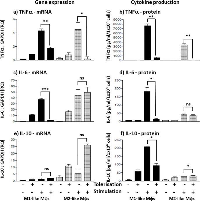
Fig 3. E.coli K12-LPS differentially suppresses Mφ subset cytokines secretion and gene expression.
M1 (bold) and M2 (shaded) Mφ subsets were pre-stimulated with 100 ng/ml K12-LPS for 24 hours prior to stimulation with 100 ng/ml K12-LPS incubated for a further 18 hours, indicated using (-) = no LPS, whereas (+) = LPS added for both pre-stimulated (tolerisation) and stimulated cells (stimulation). Gene expression, mRNA, was tested in both Mφ subsets for the expression of TNFα mRNA (a), IL-6 mRNA (c) and IL-10 mRNA (e), where the mRNA level is expressed as fold change (RQ) using GAPDH as reference gene and resting cells as a calibrator sample, as described in [16] using 2-ΔΔct method. Data displayed for gene expression is a representative experiment with duplicate samples for n = 3 replicate experiments. Cytokine production was measured by sandwich ELISA and presented as the mean secretion ± SD in pg/ml for TNFα (b), IL-6 (d) and IL-10 (f). Data displayed is representative of triplicate samples for n = 3 replicate experiments. Significant effects on suppression compared to the untolerised LPS stimulation control for the specified Mφ subset are indicated as * p < 0.05, ** p < 0.01, ***P < 0.001 and ns = not significant.

