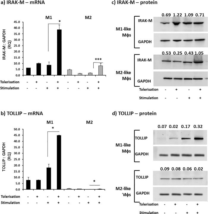
Fig 5. Endotoxin-tolerisation of Mφ subsets differentially regulates IRAK-M and Tollip.
M1 (bold) and M2 (shaded) Mφ subsets were pre-stimulated with 100 ng/ml K12-LPS for 24 hours prior to stimulation with 100 ng/ml K12-LPS incubated for a further 18 hours, indicated using (-) = no LPS, whereas (+) = LPS added for both pre-stimulated (tolerisation) and stimulated cells (stimulation). Gene expression, mRNA, was tested in both Mφ subsets for the expression of IRAK-M mRNA (a) and Tollip mRNA (b), where the mRNA level is expressed as fold change (RQ) using GAPDH as reference gene and resting cells as a calibrator sample, as described in [16) using 2-ΔΔct method. Data displayed for gene expression is a representative experiment with duplicate samples of n = 3 replicate experiments. Significant effects on suppression (+/+) compared to the untolerised LPS stimulation control (-/+) for the specified Mφ subset are indicated as * P<0.05, *** P<0.001. IRAK-M (c) and Tollip (d) protein was detected by western blotting and levels of GAPDH served as internal controls. The relative band density ratio of IRAK-M and Tollip protein compared to the GAPDH house-keeping protein loading control is indicated numerically above the appropriate sample detected on the blot. Data displayed is a representative blot of n = 3 independent replicate experiments.

