. 2019 May 23;8:e42325. doi: 10.7554/eLife.42325

Figure 1. High resolution structural images of five adult female macaques of similar age and weight.

The large bilateral lesions of monkey S to the occipital cortex are clearly visible. LGN (white arrow) and pulvinar (posterior and dorsal to LGN) are shown in parasagittal section (with high magnification inset) as well as in the coronal and horizontal sections.

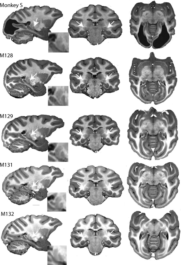
Figure 1.

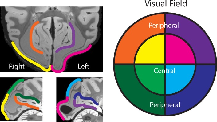
Figure 1—figure supplement 1. Visual map estimate of monkey S.

Figure 1—figure supplement 1.

Based on established neurophysiological maps of V1 retinotopy in relation to the cortical folding pattern and approximate slice position (their Figure 11, Van Essen et al., 1984), we estimated the visual field loss of monkey S. The visual map is shown on post mortem T2*-weighted images – deformations of the occipital cortical surface were post mortem. Given the extent of the loss of white matter directly underneath V1, we expect bilateral visual field loss, for the central 10° as well as for more peripheral parts, potentially around 15°−20° eccentricity.