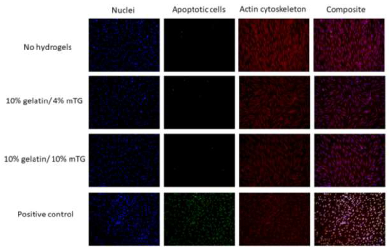
Figure 7.

Apoptosis assay. The hDFs cultured in 24 well plates were exposed to no hydrogels, 10% gelatin/4% mTG hydrogels and 10% gelatin/10% mTG hydrogels through the transwell inserts over 7 days. Cell nuclei were stained blue with DAPI, apoptotic cells were stained green with TUNEL assay and actin cytoskeleton was stained red with rhodamine-labeled phalloidin. The positive control was generated by treating the no-hydrogel samples with DNase I. The composite images were generated by merging all three channels (blue, green, red) using imageJ.
