. 2019 Apr 30;8:e43598. doi: 10.7554/eLife.43598

Figure 4. Immunofluorescent staining of PHGDH in human retina.

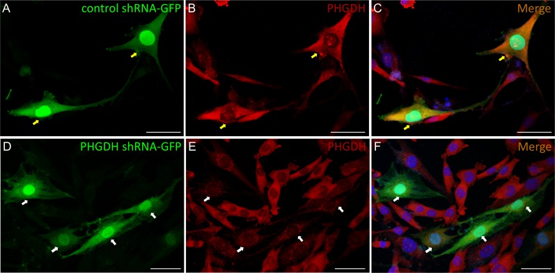
Representative images of immunofluorescence visualization of PHGDH (green) and CRALBP (red, a Müller cell marker) in human macular (A-H) and peripheral retina (I-P). E-H Field enlarged image from B (white-dotted box) in macula; M-P. Enlarged images from J (white-dotted box) in peripheral retina. The specific immunoreactivity of PHGDH antibody was verified in PHGDH knockdown in MIO-M1 cells (Figure 4—figure supplement 1).

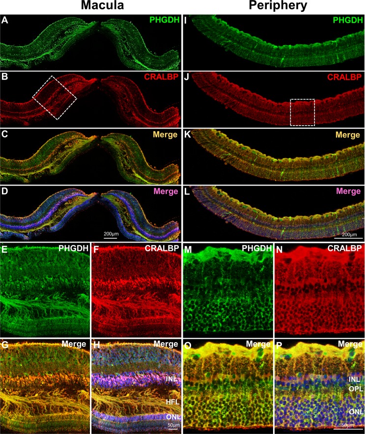
Figure 4.

Figure 4—figure supplement 1. Verification of PHGDH specific immunoreactivity by PHGDH knockdown in MIO-M1 cells.

Figure 4—figure supplement 1.

Cells expressing green fluorescence have been successfully transfected with control (A) or PHGDH (D) shRNA-GFP plasmid. PHGDH was immunostained with anti-PHGDH antibody, visualised with Alexa 594 (red) in the MIO-M1 cells (B and E). (C) Merged images of A and B showing normal PHGDH immunoreactivity in cells transfected with negative control shRNA-GFP plasmid (yellow arrows). (F) Merged images of D and E showing reduced PHGDH immunoactivity in the cells transfected with PHGDH-shRNA-GFP plasmid (white arrows). Scale bar = 50 µM.