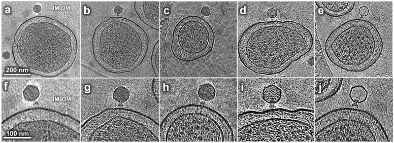
Figure 1. Tomograms reveal P22 intermediates at different stages of infection.
(a, f) A phage obliquely attached to the minicell envelope after 3 min infection; (b, g) A phage attached to the minicell surface perpendicularly after 15 min infection; (c, h) A phage attached to the minicell surface showing extended density across the cell envelope after 15 min infection; (d, i) a phage with less than a complete genome in its capsid after 30 min infection; (e, j) a phage with an apparently empty capsid after 60 min infection. IM: inner (cytoplasmic) membrane; OM: outer membrane, including O antigen. Comparable tomograms have been obtained in at least three experiments using independent preparations of minicells and phage.

