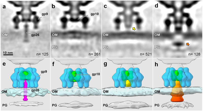
Figure 3. Intermediate structures during commitment to infection.
(a, e) An averaged structure of 125 perpendicularly adsorbed virions with the tail needle penetrating the outer membrane; (b, f) an averaged structure from 261 particles without the needle; (c, g) an averaged structure from 521 particles with an extracellular channel (yellow arrow) between the distal end of the baseplate gp10 and the outer membrane; (d, h) an averaged structure from 128 particles with channel extension and membrane cavity (orange arrow). OM: outer membrane, including O antigen; PG: peptidoglycan cell wall. All intermediates were observed in at least three experiments using independent preparations of minicells and phage.

