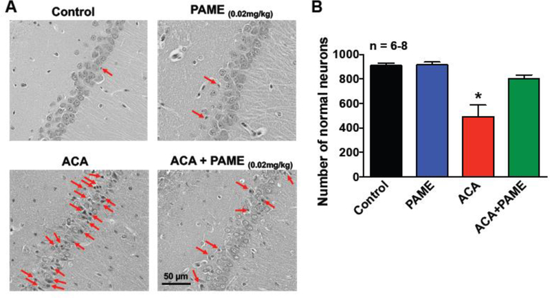
Fig. 4. Post-treatment of PAME enhanced neuronal survival in the CA1 region of the hippocampus after ACA.

(A) Representative images of H&E in the CA1 region of the hippocampus. Rats received an IP bolus injection of PAME (0.02 mg/Kg) or vehicle (0.0026% ethanol) immediately after ACA. Animals were sacrificed 7 days after ACA for brain histopathology of the hippocampus. Control (no ischemia, no drug) and PAME (vehicle control) groups were performed as internal controls. A lightly stained nucleus with a dark-stained nucleolus and a red-stained cytoplasm can be observed in healthy neurons, while ischemic neurons exhibit shrunken cytoplasm and pyknotic nuclei. Arrows denote typical neuronal cell death in the CA1 region of the hippocampus. Short horizontal solid bars represent 50 µm in length in the field of view of each representative image. (B) The number of healthy neurons from the CA1 region of the hippocampus were counted and expressed in the bar graph. n number of animals used per group. *p≤0.05 indicates significantly different from all groups, evaluated by one-way ANOVA with Tukey’s post-hoc test.
