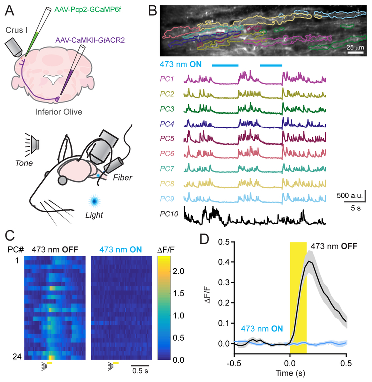
Figure 3. Dendrite-wide Ca2+ signals in PCs are completely mediated by CFs.
(A) GtACR2 was expressed in excitatory neurons of the inferior olive; dendritic Ca2+ activity was measured in GCaMP6f-expressing PCs of Crus I during sensory stimuli. Sensory cues were presented to the animal while, in a subset of trials, an optical fiber implant continuously delivered laser light to the inferior olive.
(B) Spontaneous Ca2+ activity in PCs, demarcated in the fluorescence image above, was abolished during photo-illumination of the inferior olive (λ = 473; 250 μW). An example PC (PC10) is included that was persistently active during the optogenetic stimulus.
(C) Ca2+ activity measurements for individual PCs in a field of view in response to an auditory stimulus in control or during the continuous optogenetic suppression of the inferior olive (λ = 473; 250 μW).
(D) Average Ca2+ activity transients across PC dendrites to sensory stimuli (including both auditory and visual cues) in control and with optogenetic suppression of the inferior olive. Data are mean ± SEM (n = 234 cells from 4 mice; 10 trial blocks for each condition).
See also Figure S5–S7.

