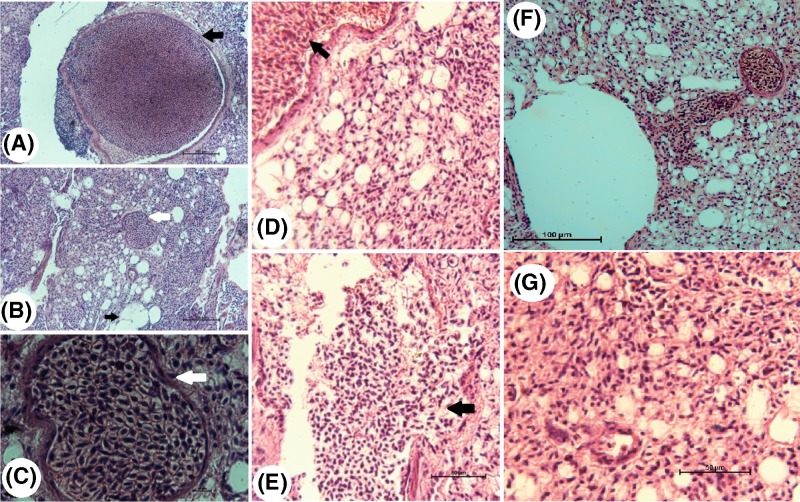
Figure 4. Photomicrographs of lung tissue sections from infected (A–E) and treated (F and G) squabs.
(A) Large schizont invading the endothelium lining of the lung tissue from squabs infected with H. columbae (black arrow, H&E, ×20), (B) small schizont (white arrow, H&E, ×20), (C) high magnification of B (white arrow, H&E, ×100), (D) schizonts in the endothelial cells of lung blood vessels (black arrow, H&E, ×40), (E) schizonts are ruptured and merozoites are disseminated in between the lung cells (black arrow, H&E, ×40), (F) schizonts were observed invading the blood vessel endothelium in squabs treated with Butalex® (H&E, ×20) and (G) schizonts were not seen in lung tissue sections of squabs treated with Eugenol (H&E, ×20).

