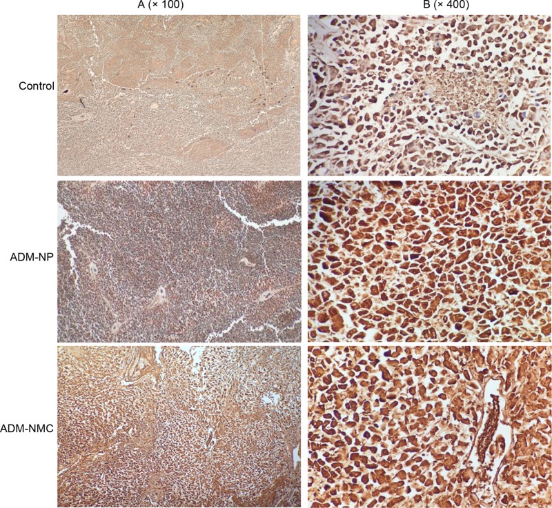
Figure 6. Immunohistochemical staining of tumor sections for Bax.
Positive staining of Bax was indicated by granular brown staining primarily located in the cytoplasm. (A) Light microscope images of the control, ADM-NP and ADM-NMC groups (magnification, ×100); scale bar, 100 μm. (B) Light microscope images of the control, ADM-NP and ADM-NMC groups (magnification, ×400); scale bar, 15 μm. ADM-NP group, adriamycin-loaded PLGA nanoparticle group; ADM-NMC, adriamycin-loaded PLGA nanoparticle microbubble complex group; Bax, B cell lymphoma-2-associated X protein; PLGA, polylactic-co-glycolic acid.

