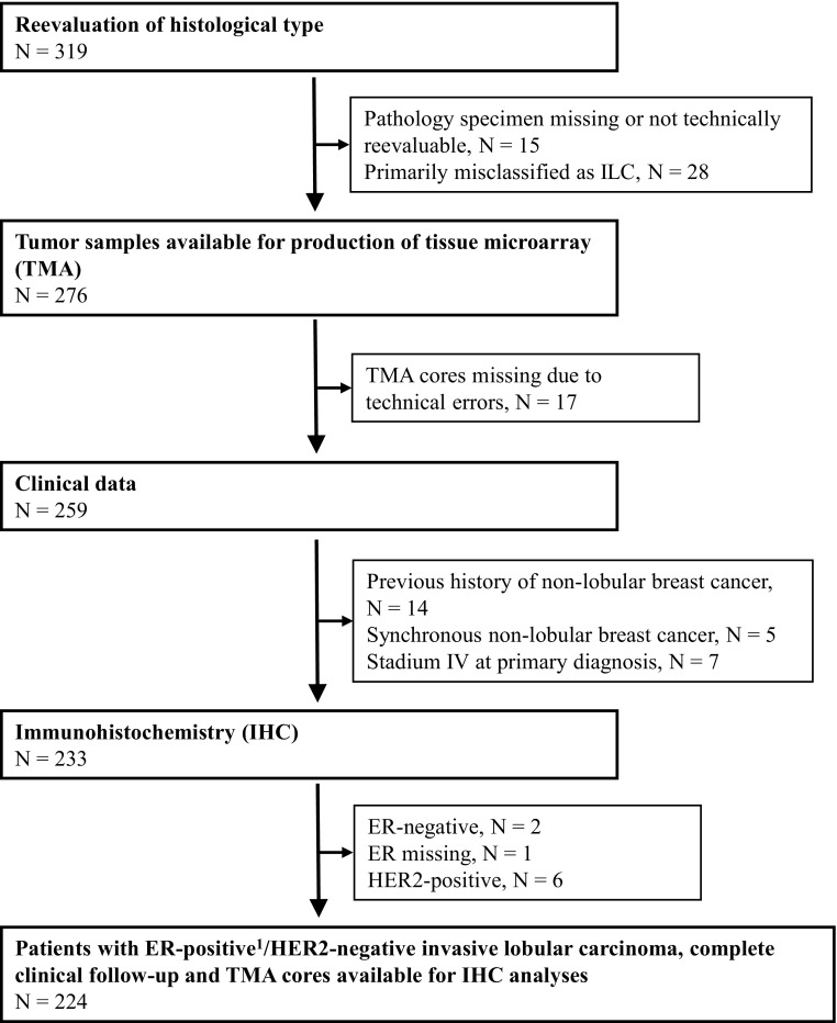
Fig. 1.
Consort diagram: Breast cancer patients with tumors primarily classified as invasive lobular carcinomas (ILC) at the Department of Pathology, Skåne University Hospital Lund and Helsingborg Hospital, (1980–1991), N = 319. ER positivity (≥ 1%) was confirmed with IHC staining on tissue microarray in N = 200 and whole tissue sections in N = 21, and with cytosol-based methods in N = 3 tumor samples

