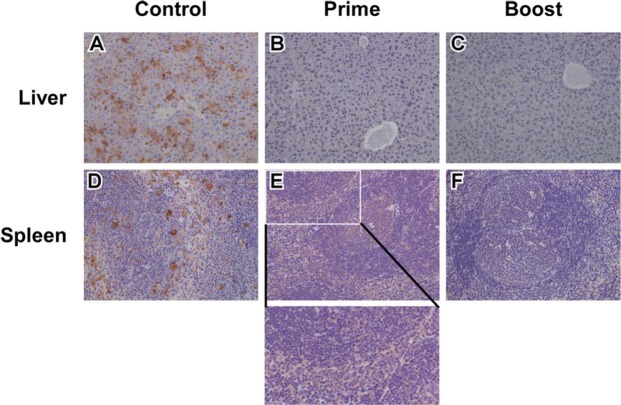
Figure 6.
Immunohistochemistry of mice tissues. Representative study endpoint liver tissue sections for the control (A), prime (B), and boost (C) cohorts with observable marked CCHFV immunolabeling of hepatocytes in the control group (A) and no observable labeling in the prime (B) or boost (C) cohorts. Representative study endpoint spleen tissue sections for the control (D), prime (E), and boost (F) cohorts with observable marked CCHFV immunolabeling in mononuclear cells (D), cytoplasmic, mild, and diffuse immunolabeling of mononuclear cells primarily in the red pulp (E, inset), and no specific CCHFV immunolabeling in the boost cohort (F). Original magnification of all panels, 20X.

