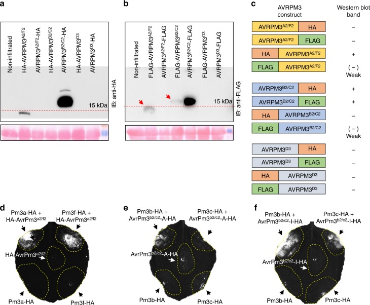
Fig. 2.
Impact of epitope fusion of the AVRPM3 protein expression and detectability. a, b Western blot detection of C and N terminal HA (a) and FLAG (b) epitope tag fusion of AVRPM3A2/F2, AVRPM3B2/C2 and AVRPM3D3 (upper panel) and Ponceau staining of the Western blot membrane (lower panel) are depicted. Uncropped western blot images are provided in a Source Data File. c Graphical summary of AVRPM3 protein tagability based on detection of the protein on a western blot, demonstrating significantly different impacts of epitope fusions based on tag position and sequence. d–f experimental assessment of HA tagged AVR variants in functional validation assays in N. benthamiana. Protein expression data is provided in Supplementary Fig. 6. HA-AVRPM3A2/F2 (d), AVRPM3B2/C2-A-HA (e) and AVRPM3B2/C2-I-HA (f), co-expressed with their respective NLRs. HR was assessed using HSR imaging 4–5 days after Agrobacterium infiltration. Results are consistent over at least two independent assays each consisting of 6–8 independent leaf replicates

