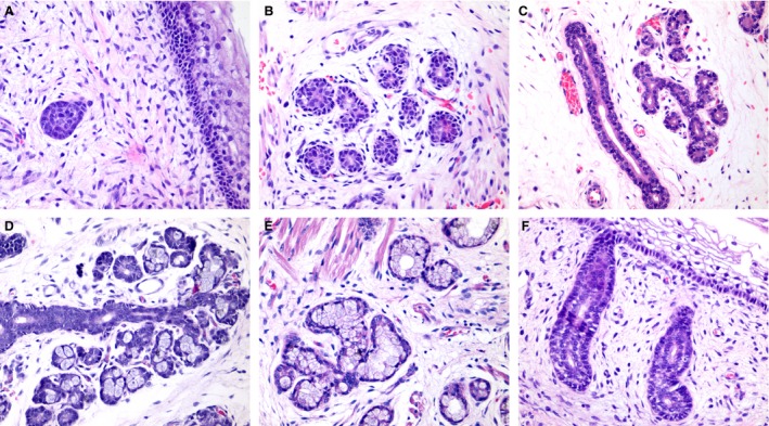
Figure 1.

Histological aspects of human salivary glands morphogenesis. (A) Initial bud stage showing a solid group of epithelial cells. (B) Pseudoglandular stage showing the early luminal opening. (C) Canalicular stage showing branched luminal structures. (D) Terminal bud with the presence of early secretory units in branched structures. (E) Well‐differentiated secretory units. (F) Excretory duct connecting to the mucosa. Magnifications A–E = 400 ×.
