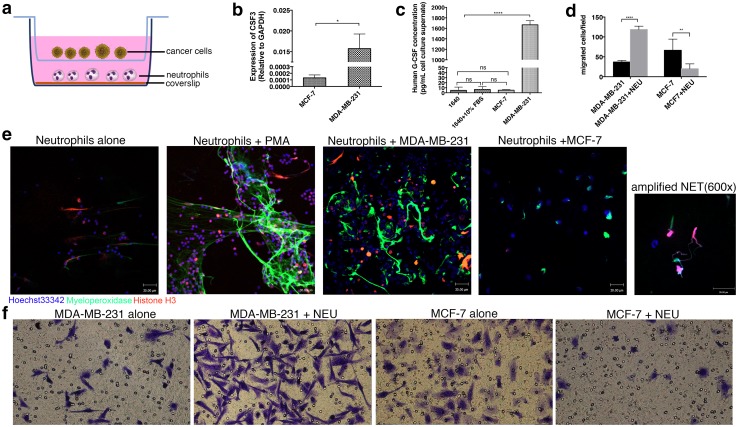
Fig. 6.
NETs formed by G-CSF high-expression MDA-MB-231 cells promote cancer cell migration. a Schematic of the migration/NET formation transwell assay. Fresh neutrophils were seeded on the coverslips layered in the lower chamber, and cancer cells were placed in the upper chamber. b and c Production counts of G-CSF in MDA-MB-231 and MCF-7 cells detected by qRT-PCR (mean ± SEM; t test) and ELISA (mean ± SEM; ANOVA) assays. d and f Neutrophils increased the motility of MDA-MB-231 cells but not MCF-7 cells (mean ± SEM; two-way ANOVA with multiple comparisons). e Unstimulated neutrophils and neutrophils primed by PMA, MDA-MB-231 cells and MCF-7 cells showed different statuses (immunostaining for myeloperoxidase and histone H3). PMA and MDA-MB-231 cells prime neutrophils into NETs. Significant differences are indicated with *, whereas nonsignificant differences are indicated with ns. *p < 0.05; **p < 0.01; ***p < 0.001; ****p < 0.0001

