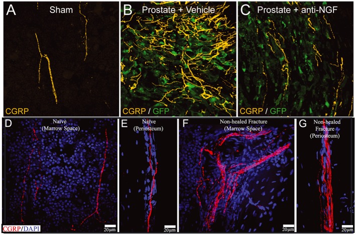
Figure 4.

Images of nerve sprouting in malignant and non‐malignant bone pain. As seen in (A) sensory nerve fibres (bright yellow) in the normal bone marrow have a regular and linear appearance. Following invasion of the bone marrow (B) by prostate cancer cells (green) there is marked increase in the density due to the ectopic sprouting of sensory nerve fibres. Prior administration of anti‐nerve growth factor (NGF) before tumour invasion of the bone largely blocks this ectopic nerve sprouting (C). Similar sprouting of sensory nerve fibres is also observed in non‐malignant pain states following injury of bone. (D, E) Sensory nerve fibres (red) in the normal marrow and periosteum respectively. (F, G) Increased sensory innervation of the marrow (F) and periosteum (G) in a bone at 6 months postfracture when effective bone healing has not occurred. Abbreviations: anti‐NGF, a monoclonal antibody that binds to nerve growth factor (NGF); CGRP, calcitonin gene related peptide (labels small diameter sensory nerve fibers); DAPI, 4′,6‐diamidino‐2‐phenylindole (a counterstain that labels the nucleus of all cells); GFP, green fluorescent protein that is expressed by prostate cancer cells
