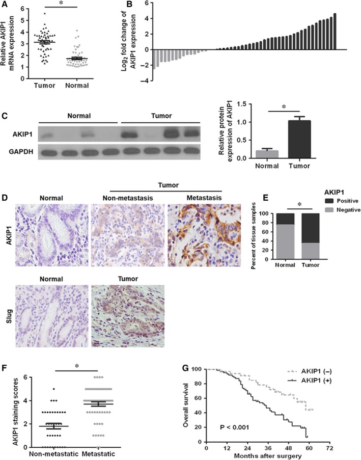
Figure 1.

Relative A‐kinase‐interacting protein 1 (AKIP1) expression in gastric cancer (GC) tissues and its clinical significance. (A, B) Quantitative real‐time PCR analysis of AKIP1 mRNA expression in GC tumour tissues and corresponding normal tissues. (C) Western blot analysis of AKIP1 protein expression in GC tumour tissues and matched normal tissues. (D) Representative immunohistochemistry images of AKIP1 and Slug in GC tissues and adjacent normal tissues. (E) Quantitative assessment of AKIP1 expression in tumour tissues and matched normal tissues in accordance with staining scores. (F) Scatterplot of the staining scores of AKIP1 expression in patients without or with metastasis. (G) Kaplan‐Meier survival curve for GC patients with AKIP1 expression. *P < 0.05
